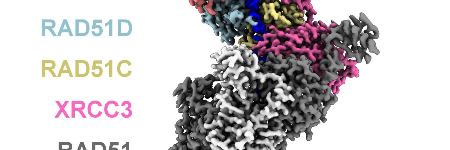
Publication highlights

Go inside our research

Explore a selection of research case studies from the past five years.

Read now
A Crick researcher reading a scientific paper on a screen.

Intro

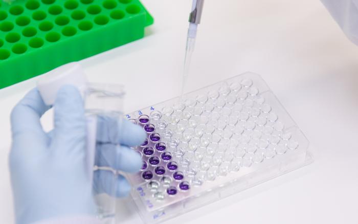
Researchers at the Crick are tackling the big questions about human health and disease, and new findings are published every week.

Our faculty have picked some of the most significant papers published by Crick scientists, all of which are freely available thanks to our open science policy.

Teams

Highlights

Cytokines

A balancing act: preventing an overactive immune system

Researchers at the Crick, University of Cambridge, Sanquin and the NOVA University investigated how T cells switch off immune functions as quickly as they are switched on, looking at two mRNA shutdown signals: AU-rich elements (long stretches of nucleotides that signal to other proteins to degrade the mRNA) and m6a methylation (adding chemical red flags to mRNAs to mark them for removal). They mapped all m6a methylation sites in human T cells before and after activation, observing that m6a methylation doesn't happen randomly, but often takes place near AU-rich elements. When these two signals occurred close together, the mRNA rapidly degraded, referred to as 'meta-unstable'. This system allows the immune system to keep the balance between under and overactivation.

Meta-unstable mRNAs in activated CD8+ T cells are defined by interlinked AU-rich elements and m6A mRNA methylation

Published in Nature Communications

Published

Synthetic sugars

Sweet signals: tracking crucial cell messengers for the first time

Researchers at the Crick and Imperial College report a method to characterise and track sugar-coated cell sensors called proteoglycans using click chemistry. Through a 'bump and hole' engineering technique, they modified a hole in an enzyme and a bump in a sugar, to alter an enzyme that glues the two together so it accepts a bumped version of the sugar. This modified sugar contains a chemical tag which means it can be traced using click chemistry, such as attaching a fluorescent molecule to 'see' the molecule by imaging, or a molecule acting like an anchor to isolate and further study it. In the future, these molecules could be tagged and tracked in different contexts, or proteoglycan function could be altered by replacing the sugar chain with a different biological or synthetic molecule.

Xylosyltransferase engineering to manipulate proteoglycans in mammalian cells

Published in Nature Chemical Biology

Published

Cell death programmes

Unravelling a cell death programme evaded by half of all cancers

When normal cells become cancer cells, they undergo a series of genetic changes that allow them to divide indefinitely. One such change involves the loss of a protein called Schlafen 11 (SLFN11), which occurs in half of all cancers. SLFN11 activity results in programmed cell death in response to damaged DNA, which naturally occurs during cancer cell transformation. Thus, loss of SLFN11 renders cancer cells immune to DNA damage and resistant to wide range of chemotherapies currently used in the clinic. However, how damaged DNA activates SLFN11 to cause programmed cell death is not known. Here, researchers at the Crick have uncovered what cellular processes lead to a specific type of DNA damage that activates SLFN11 and programmed cell death. This work provides insight as to why half of all cancers lose SLFN11 in response to naturally occurring DNA damage.

RPA exhaustion activates SLFN11 to eliminate cells with heightened replication stress

Published in Nature Cell Biology

Published

HeLa cells with and without f-actin antigen

Lifting cancer’s invisibility cloak

Researchers at the Crick investigated whether dendritic cells detect dead cancer cells via a receptor called DNGR-1, which detects F-actin. Looking at mice with and without the DNGR-1 receptor that had been exposed to carcinogens, they found that mice without DNGR-1 developed tumours significantly earlier and to a greater extent. Next, the team examined whether certain cancer mutations were more likely to be found in the tumours of mice without DNGR-1. They reported an increase in mutations in proteins that bind to the F-actin scaffold. This may be because, in mice with DNGR-1, mutations in these proteins are highlighted as a red flag for the immune system. Without DNGR-1, there's less evolutionary pressure for cancer cells to get rid of them.

Cross-presentation of dead cell-associated antigens shapes the neoantigenic landscape of tumor immunity

Published in Nature Immunology

Published

Lung-on-chip

Built to breathe: mini ‘lungs’ recreate individual response to infection

Researchers at the Crick and AlveoliX have developed the first human 'lung-on-chip' model using stem cells taken from only one person. The team produced type I and II alveolar epithelial cells and vascular endothelial cells from human-induced pluripotent stem cells. These epithelial and endothelial cells are separatley grown on the top and bottom of a very thin membrane in a device to recreate an air sac barrier, which experience rhythmic three-dimensional stretching forces on the recreated air sac barrier, mimicking the motion of breathing. The scientists then added macrophages into the chip, before adding TB bacteria. In the chips infected with TB, the team reported large macrophage clusters containing a group of dead macrophages in a necrotic core.

Autologous human iPSC-derived alveolus-on-chip reveals early pathological events of Mycobacterium tuberculosis infection

Published in Science advances

Published

Fly wing growth

Oxygen availability constrains growth during development

Growth is a key feature of development, but animals, organs and tissues must know when to stop growing. Researchers at the Crick have shown that the sac-like structures that give rise to fly wings do not stop growing abruptly. Instead, growth slows down over the course of days. Measurements of global gene activity during growth deceleration suggest that, as the primordium expands, it becomes increasingly hypoxic. Decreasing oxygen availability, perhaps due to inefficient import as tissue size increases, was confirmed with new genetic sensors of cellular oxygen. This study uncovers a feedback loop whereby growth (and increasing tissue size) leads to hypoxia, which in turn dampens growth to ensure that oxygen demand does not overwhelm dwindling supplies.

HIF-1α-mediated feedback prevents TOR signalling from depleting oxygen supply and triggering stress during normal development

Published in Nature Communications

Published

Neural tube and somites

Uncovering early embryonic communications using new stem cell model

Researchers at the Crick have produced a new embryo model that self-organises around ten somites alongside a single neural tube, mirroring aspects of human embryos at 28 to 35 days after fertilisation. As the models don't contain a notochord, the team introduced signals that would have originally come from a notochord, and observed a shift in cell fates. They also saw spontaneous patterning in the neurla tube, showing it was developing into different identieis depending on the cell's location. This suggested that the somites and the neural tube were in close communication. The team confirmed that increased retinoic acid signalling in specific somite regions was likely due to signalling to the neural tube, allowing spontaneous patterning. This crosstalk helps prompt regional identities and may be important for later maturation to neuronal or skeletal tissues.

Modelling co-development between the somites and neural tube in human trunk-like structures

Published in Nature Cell Biology

Published

We are very interested in molecules called RNAs, which are produced when particular sections of DNA are ‘read’ and are thought to be involved in controlling gene activity and differentiation.

"Canary in a coal mine" for mitochondrial dysfunction

Mitochondria are the cell’s powerhouses and are essential for organismal health. When they malfunction, proteins meant to enter them can accumulate outside and act as distress signals, alerting the cell to potential damage. Researchers at the University of British Columbia, in collaboration with colleagues at the Crick, discovered that a small region of a mitochondrial protein plays a key role in activating a protective program that promotes mitochondrial recovery. Under normal conditions, this region enables the protein to enter mitochondria, but when blocked, it switches roles to signal stress. This finding reveals a natural “canary in a coal mine” for mitochondrial dysfunction and opens new possibilities for treating neurodegenerative and other mitochondria-related diseases.

A direct role for a mitochondrial targeting sequence in signalling stress

Published in Nature

Published

Astrocytes

Uncovering early hypoxic stress in ALS astrocytes

Researchers at the Crick and UCL have shown that reported that astrocytes show signs of hypoxic stress long before neurons begin to die in ALS. Using stem cells from patients to generate astrocytes carrying ALS-linked mutations in a gene called VCP, which is linked to inherited forms of ALS, the team showed that astrocytes exhibited clear signs of 'pseudo-hypoxia'. This meant they had switched on a low-oxygen response despite being in normal oxygen conditions. This was driven by HIF-1a, a master regulator of how cells respond to oxygen. Instead of being degraded under normal conditions, it had accumulated in the nucleus and activated genes involved in metabolism, energy production and stress responses. As a result ALS astrocytes showed mitochondrial dysfunction and a reduced ability to support motor neurons. This is particularly exhibited as an inability to correct the mislocalisation of RNA-binding proteins, a well-known molecular hallmark of ALS, compromising neuron survival.

Hypoxic stress is an early pathogenic event in human VCP-mutant ALS astrocytes

Published in Stem Cell Reports

Published

E.coli

Researchers rescue antibiotics from resistance using phototherapy

Researchers at the Crick and King's College London have used phototherapy to inhibit a protein in E. Coli bacteria that makes them resistant to antibiotics. They designed a new chemical tool, Ru1, composed of a light-activated ruthenium metal complex attached to an organic ligand that binds to NDM-1, an enzyme in drug-resistant bacteria that breaks down common beta-lactam antibiotics like penicillin. When exposed to blue light, the metal complex produces reactive oxygen species that cause damage to NDM-1, preventing it from binding and destroying an antibiotic. They showed that Ru1 can boost the activity of meropenem antibiotic against E. Coli by 53 times, without showing toxicity to human cells.

Light-activated metal-dependent protein degradation: A heterobifunctional ruthenium(II) photosensitizer targeting New Delhi metallo-β-lactamase 1

Published in Journal of the American Chemical Society

Published

Volume EM and X-ray imaging

X-ray imaging captures the brain’s intricate connections

Researchers at the Crick and the Paul Scherrer Institute have developed a new imaging protocol to capture mouse brain cell connections in precise detail. Building on standard volume EM sample preparation protocols, they tested a new step - embedding the stained tissue using a resin developed in the nuclear and aerospace industries to protect against radiation. The samples were then imaged using X-rays in a synchrotron. The resulting images, produced using a specific type of X-ray imaging called X-ray ptychography, reached a resolution of 38nm. This was enough to show multiple elements of the mouse brain circuitry, including synapses, dendrites and axons.

Nondestructive X-ray tomography of brain tissue ultrastructure

Published in Nature Methods

Published

Cave where wolf remains were found

Ancient wolves on remote Baltic Sea island reveal link to prehistoric humans

Researchers at the Crick, Stockholm University, the University of Aberdeen and the University of East Anglia analysed two 3,000-5,000-year-old wolf remains found in the Stora Förvar cave on the Swedish island of Stora Karlsö. A small, isolated island, Stora Karlsö has no native land mammals, meaning that any animals found must have been brought there by people. Genomic analysis of the two canid remains confirmed they were wolves, not dogs, with no evidence of dog ancestry. However, they exhibited several traits typically associated with life alongside humans, such as a diet aligned with the humans on the island, a size smaller than typical mainland wolves, and low genetic diversity, a common result of isolation or controlled breeding. The finding challenges the conventional understanding of wolf-human dynamics and the process of dog domestication.

Gray wolves in an anthropogenic context on a small island in prehistoric Scandinavia

Published in Proceedings of the National Academy of Sciences of the United States of America

Published

Giant cancer cells in sarcomas

Giant cancer cell dynamics in sarcomas

Researchers at the Crick examined unusually large and abnormal cells called polyploid giant cancer cells (PGCCs) in ten pleomorphic sarcomas, types of soft-tissue cancers known to be highly aggressive and genetically complex. Using advanced single-cell DNA sequencing, they analysed the genetic material of individual PGCCs to see how they differ from the rest of the tumour. They found that PGCCs were scattered randomly rather than forming groups in the tumour, suggesting that they arise spontaneously. They appeared to come from the main tumour cell population but had more genetic variation and many had signs of chromosomal instability. Chromothripis, where chromosomes shatter and reassemble in a chaotic pattern, was frequently seen in PGCCs. This ongoing genomic reshaping may explain why pleomorphic sarcomas often behave aggressively and are difficult to treat.

Profiling the genomic landscape and evolutionary history of polyploid giant cancer cells in undifferentiated pleomorphic sarcomas

Published in Cancer Letters

Published

Lipid envelopes on TB bacteria

Scavenger hunt: how TB bacteria overcome nutrient scarcity

Researchers at the Crick have discovered that Mtb, the bacterium causing tuberculosis (TB), alters its outermost layer, its lipid cell envelope, when it encounters low phosphate conditions. This allows it to survive inside human immune cells, where phosphate is restricted. It can scavenge phosphate from human lipids (fats), which are present in the lungs, allowing the bacteria to grow when no other source of phosphate is present. These findings demonstrate a method that Mtb employs to overcome the human host’s attempts to restrict its growth. The replacement lipids produced
when phosphate is restricted therefore represent new drug targets for the treatment of TB. Additionally, vaccines that target TB via its lipids should take into account the particular lipids present when the cell is phosphate starved, as demonstrated here.

Mycobacterium tuberculosis overcomes phosphate starvation by extensively remodelling its lipidome with phosphorus-free lipids

Published in Nature Communications

Published

firebrat and fruit fly

When evolution took flight

Researchers at the Crick have identified a signalling feedback loop which they think may have been vital to the evolution of insect wings and therefore flight. They found that, as concentrations of a morphogen called Dpp decrease across the wing tissue, another molecule called Brinker forms a reverse gradient. The Brinker gene is repressed by Dpp and is therefore increasingly expressed as the Dpp signal becomes weaker. They then found that Brinker is only found in insects and not in closely related crustaceans, and that it is found in a wingless insect called a firebrat, but doesn't form a gradient and is as yet unconnected to the Dpp signal transduction. This suggests that the Brinker-mediated feedback circuit may have been an evolutionary innovation of winged insects.

A genetic circuit that extends the useful range of a BMP morphogen arose alongside insect wing evolution

Published in Current Biology

Published

Artificial ion channels

Artificial ion channel recreates membrane protein functions

Natural ion channels of biology allow cells to communicate, transfer nerve impulses, trigger sensations, and cellular processes. Biology has a variety of highly effective channels, but creating new, orthogonal systems is challenging. Researchers at the Crick have designed a system able to span a lipid bilayer, with a single internal channel, which allows the passage of certain anions and cations. They can control its activity using three biorthogonal handles - light, pH, and presence of a 'guest' molecule, which blocks the channel. This allows them to formulate a molecular logic gate, achieving a simple analogy of the complex functions of biological transmembrane proteins.

Triply responsive control of ion transport with an artificial channel creates a switchable AND to OR logic gate

Published in Angewandte Chemie International Edition

Published

Macrophages with and without ARPC5

How weakness in cell structure affects the host-microbiome relationship

Children born with mutations in the ARPC5 protein, which is part of the internal cytoskeleton, experience immunodeficiency and a high risk of sepsis. Researchers at the Crick investigated immune system function in mice with and without ARPC5 mutations, observing inflammation in adult mice with ARPC5 deficiency that mirrored that in humans. They showed that this was due to a big change in bacterial composition in the gut after weaning, triggering intestinal inflammation, as giving antibiotics to ARPC5-deficient mice at a critical four-week time point fully prevented the disease from developing. Finally, the team showed that macrophages with ARPC5 mutations had lost their usual shape and could no longer kill bacteria effectively, leading to an overwhelming response to the microbiome.

Branched actin networks mediate macrophage-dependent host-microbiota homeostasis

Published in Science

Published

RAD51 complex

Capping RAD51 filaments to protect genome stability

Our cells rely on DNA repair systems to prevent genome instability and cancer. One of the most accurate is homologous recombination, driven by RAD51 and assisted by five RAD51-like proteins whose roles were unclear. Using cryo-electron microscopy, biochemistry and single molecule analyses, Crick researchers show that these proteins assemble into two distinct complexes. The RAD51B complex helps initiate repair by assembling RAD51 filaments, while the XRCC3 complex plays the more ancient and conserved role: capping and stabilising RAD51 filament ends and promoting DNA strand pairing. This work uncovers a fundamental mechanism for genome protection and clarifies how mutations in RAD51-like genes contribute to cancer.

Cryo-electron microscopy visualization of RAD51 filament assembly and end-capping by XRCC3-RAD51C-RAD51D-XRCC2

Published in Science

Published

Limb malformation in PRKCA mutations

Discovery reveals new understanding of cancer-driving proteins in rare brain tumours and beyond

Scientists at the Crick and Barts Cancer Institute (Queen Mary University of London) have discovered that a single letter change in the PRKCA gene drives a rare and hard-to-treat brain cancer, chordoid glioma. The PRKCA gene contains instructions for making a protein called protein kinase C alpha (PKCa). Until now, many believed blocking kinases would be useful for treating cancer, but in this study the team discovered that the mutation in PRKCA blocks the kinase but paradoxically drives tumour growth. This was because it became locked in a shape that allowed it to promote cancer cell growth signalling and because it interacted with epigenetic regulators in a way that promoted cancer growth.

The chordoid glioma PRKCA D463H mutation is a kinase inactive, gain-of-function allele that induces early-onset chondrosarcoma in mice

Published in Science Signaling

Published

Dendritic cells and phagosomes

Dendritic cell receptors deliver messages about immune threats quietly

A subset of dendritic cells, type 1 conventional dendritic cells (cDC1s), plays a key role in recognising material from dead or damaged cells and showing fragments of that material to killer T cells in a process known as cross-presentation. This is critical for defence against some viruses and cancer. This study uncovers how one cDC1 receptor, DNGR-1, promotes cross-presentation of antigens from dead cells while keeping the cell otherwise 'quiet'. The team discovered that this behaviour depends on a single amino acid within the receptor. Changing this amino acid switches DNGR-1 into an activating receptor, but at the cost of losing cross-presentation efficiency. The findings reveal that DNGR-1 has evolved to prioritise information gathering from dead cells over full immune activation, helping the body learn from self-damage without triggering harmful inflammation.

DNGR-1 signalling limits dendritic cell activation for optimal antigen cross-presentation

Published in EMBO Journal

Published